Our Philosophy
Anatomy Standard is a revival and evolution of the classical tradition of anatomy illustration.
Our visualizations of the structure of a human body are a combination of art qualities of classical anatomy illustration and the latest advances in the science of medicine and technology.
Best tradition of anatomy illustration
The classical – early modern – anatomists are characterized by the strong will to understand the structure of the human body and naturalistically visualize it.
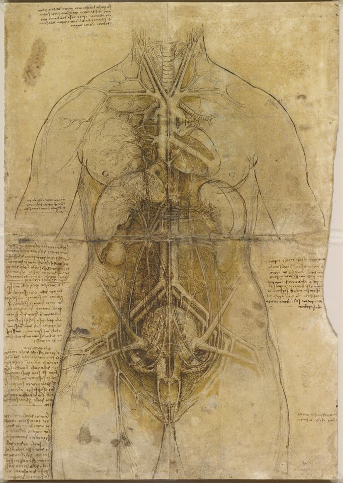
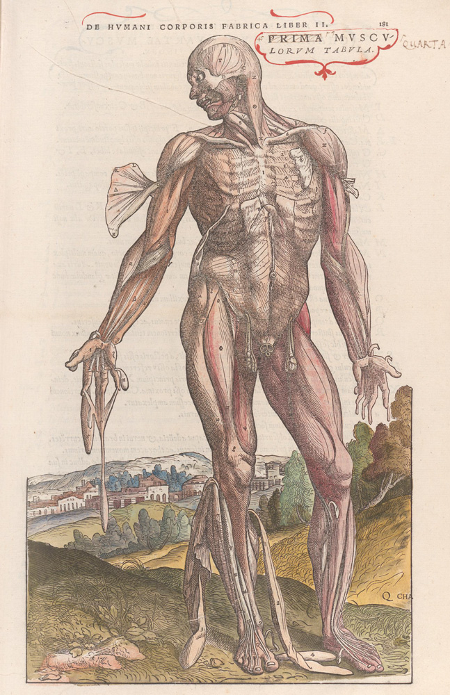
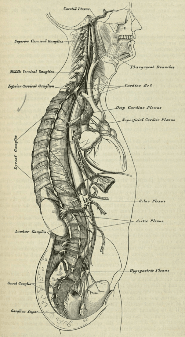
Typical examples of these efforts are anatomical drawings of Leonardo da Vinci (1452–1519), illustrations of Andreas Vesalius (1514–1564), or Charles Landseer (1799–1879). Their artworks have the right balance between realism and simplification necessary to improve the perception of the illustration, and in many aspects, are anatomically accurate.


This tradition is a base for classical anatomy works like those created by Jean Mark Bourgery (1797–1849) and Henry Gray (1827–1861) in the 19th century, Frank Netter (1906–1991) and Robert Acland (1941–2016) – in the 20th century. These books have detailed illustrations, a short, clear, and precise description of anatomical terms.

We are fascinated and inspired by these examples and many of the qualities we find in them we wish to incorporate into our works, among them elements of artistic expression like excellent contrast in shading, clear shapes and silhouettes, fine level of detail.
It is not easy to do; it demands full dedication. Nevertheless, we think that excellent results that we achieve with this approach are worth the invested effort and resources.
Anatomy Standard 3D human
Advances in the fields of 3D computer graphics and medicine – ultrasound, computed tomography, magnetic resonance – have given us a way to push the quality of anatomy visualization to the next level.
Our goal is to upkeep the best traditions of anatomy illustration and supplement them with the possibilities of modern technology to develop a 3D model of human anatomy.
Using DICOM* data sets, we will gradually build a model of healthy and physically well developed 25–35-year-old male with no prominent degenerative aging-related signs and with a full range of joint motion. To achieve this, we modify the shape, size, and position of the 3D anatomical structures guided by morphometric studies and animate the model within a particular range of motion, following biomechanics studies.
Our model of human anatomy is a base for a variety of teaching and learning resources – from illustrations seen on this website to interactive real-time applications that are currently in development.
Following our philosophy, we do our work and share the results on our website and social media. We are honored when Anatomy Standard is noticed and found useful.
Image policy
All images on our site are free to use for education and non-commercial purposes. The only thing we ask – please do not remove Anatomy Standard watermarks or logos and provide a link to our website when reposting/presenting them.
Any commercial use of our work is strictly prohibited.
Patients' DICOM data are used only after the involved persons' sign an informed consent approved by a local ethics committee.