search

Masticatory Muscles

The muscles directly involved in the chewing process, which includes elevation, depression, protrusion, retrusion, and lateral movement of the mandible, are referred to as "masticatory" muscles (from the Latin masticatio – chewing, mastication). Functionally, these muscles work in opposition to the muscles responsible for opening the mouth. Mandibular movement is more complex than it may seem. To observe the muscles of mastication in action, please explore our "Skull, Teeth & TMJ" application.

We have four pairs of masticatory muscles. They all originate from the 1st pharyngeal arch during embryonic development and are therefore innervated by the trigeminal nerve*.


  • *Toro–Tobon S, Manrique M, et al. Pharyngeal Arches, Chapter 1: Normal Development and Derivatives. J Craniofac Surg, 2023, 34(7):2237–2241.

  • manicula
    • The list to remember:
    • Masseter
    • Temporal
    • Medial pterygoid
    • Lateral pteygoid

    Skull and cervical spine with the masticatory muscles and associated ligaments. Anterior and oblique anterior aspect.
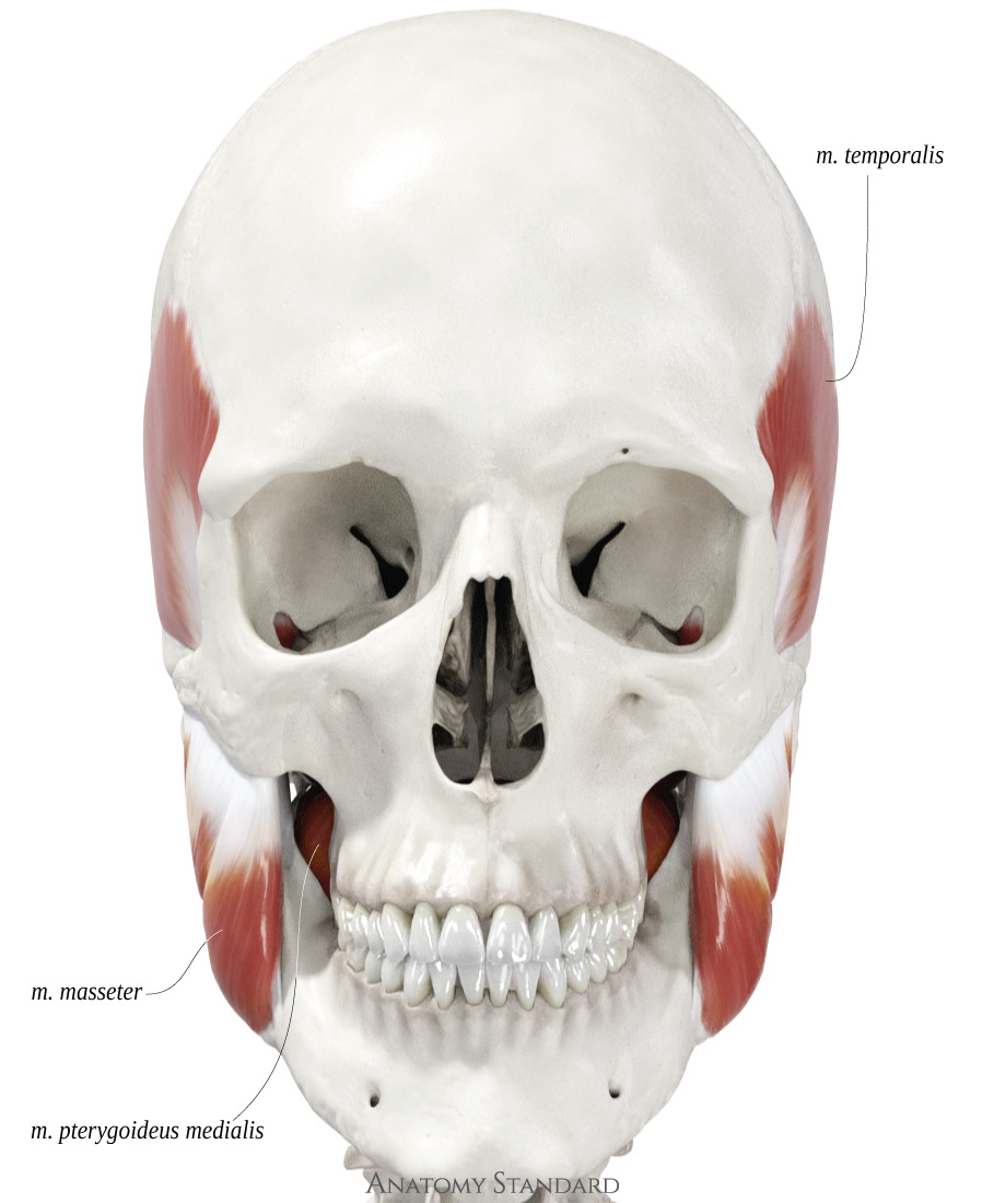
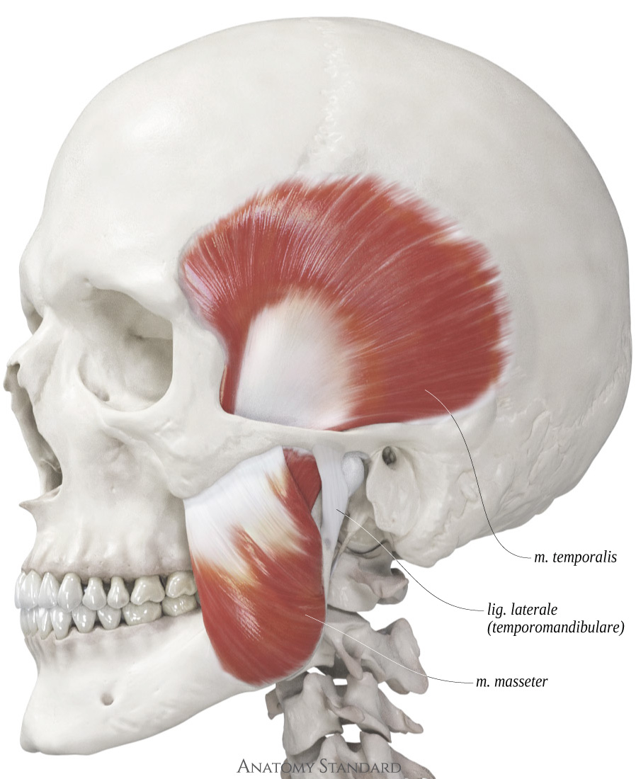
    The masseter and temporalis muscles are located superficially, so their movements are visible during active chewing.

     Skull with the masticatory muscles and associated ligaments. Posterior oblique aspect.
    Two other masticatory muscles, the medial and lateral pterygoid, are situated on the inner side of the mandible, and their presence is less evident. Nevertheless, the pterygoid muscles play a crucial role in enabling the full range of lower jaw motion and in coordinating the control of the articular disc's position in the temporomandibular joint.

    Masseter


    The masseter is the most prominent masticatory muscle. It consists of two clearly defined parts with differently directed fibers1. Some muscle fibers attach to the temporomandibular joint capsule, so the masseter muscle participates in controlling the position of articular disc. Although some authors reveal three parts2 of masseter muscle or propose more complex classification3, here we will adhere to the latest official Anatomical Terminology.


  • 1 Schumacher G. Funktionelle Morphologie der Kaumuskulatur. Fischer, Jena, 1961.
  • 2 Mezey S, Müller-Gerbl M, et al. The human masseter muscle revisited: first description of iths coronoid part. Ann Anat, 2022, Feb:240:151879.
  • 3 Brunel G, Haddioui A, et al. General organization of the human intra-masseteric aponeuroses: changes with ageing. Surg Radiol Anat, 2003, 25:270–283.
  • Icon of crossfade image Portions of the masseter muscle. Deep and superficial parts of the masseter projected on the skull. Anterolateral view
    Masseter muscle on the skull. Anterolateral view Parts of the masseter muscle projected on the skull. Anterolateral view
    Overview of the masseter
  • Superficial part:
  • Origin: zygomatic arch, mostly on the zygomatic bone
  • Insertion: angle of mandible (masseteric tuberosity)
  • Function: elevate (close) lower jaw.

  • Deep part:
  • Origin: zygomatic arch, mostly on the temporal bone & the capsule of the temporomandibular joint
  • Insertion: ramus of mandible, mostly above the masseteric tuberosity
  • Function: elevate (close) lower jaw. Lateral stabilization of the discocapsular system

  • Icon of crossfade image Deep part of the masseter muscle and the temporal muscle. Projection of origins and insertions on the skull. Oblique inferior view
    Pars profunda musculi masseteris et musculus temporalis. Inferior oblique view. Origins and insertions of the deep part of masseter and the temporal muscle. Oblique inferior aspect
    Skull with the deep part of the masseter and the temporal muscle. Oblique inferior view.
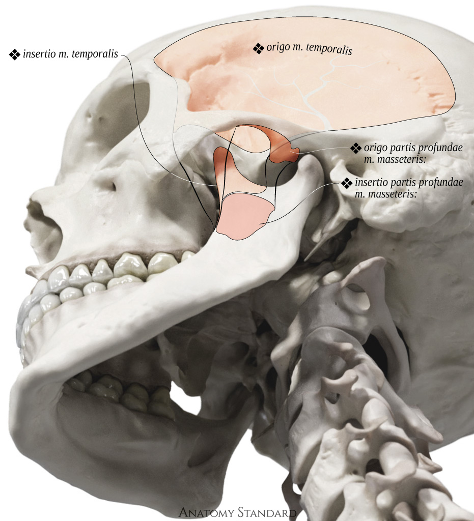
    It is worth to mention that fiber direction is similar between neibourgh deep part of the masseter and the temporal muscle. The deep part of the masseter is a kind of transition between the superficial part of masseter and temporal.
    This view also demonstrate the limited connection of the deep masseter to the capsule of the temporomandibular joint.

    Temporal muscle


    The temporal muscle, in terms of volume, is the largest among the masticatory muscles1. When it works in conjunction with the masseter muscle, these two muscles are primarily responsible for producing biting force2. Muscle fibers are derived from all the bones of the neurocranium, with the exception of the occipital and ethmoid bones. This arrangement gives rise to the distinctive fan-shaped structure of the temporal muscle.


  • 1 Takahashi M, Yamaguchi S, et al. Contribution of each masticatory muscle to the bite force determined by MRI using a novel metal-free bite force gauge and an index of total muscle activity. J Magn Reson Imaging, 2016, 44(4):804–813.
  • 2 She X, Sun S, et al. Sexual dimorphisms in three-dimensional masticatory muscle attachment morphometry regulates temporomandibular joint mechanics. J Biomech, 2021, 20:126:110623.
  • Icon of crossfade image Portions of the masseter muscle. Deep and superficial parts of the masseter projected on the skull. Anterolateral view
    Temporal muscle with skull. Side view Origin and insertion of the temporal muscle. Lateral view
    Overview of the temporal muscle
  • Origin: temporal fossa
  • Insertion: coronoid process of the mandible, pterygomandibular raphe
  • Function: elevate (close) lower jaw. Dorsal fibers are involved in the retrusion or backward movement of the mandible

  • The temporal muscle and related skull bones. Anterior, oblique anterior and oblique posterior view Icon of crossfade image
    The temporal muscle and related skull bones. Anterior aspect Origin and insertion of the temporal muscle and related skull bones. Frontal view
    The temporal muscle and related skull bones. Anterior oblique aspect Origin and insertion of the temporal muscle and related skull bones. Anterior oblique view
    The temporal muscle and related skull bones. Posterior oblique aspect Origin and insertion of the temporal muscle and related skull bones. Posterior oblique view
    The images illustrates only the skull bones that serve as the origin or insertion points of the temporal muscle. Anterior, lateral and posterior oblique view.
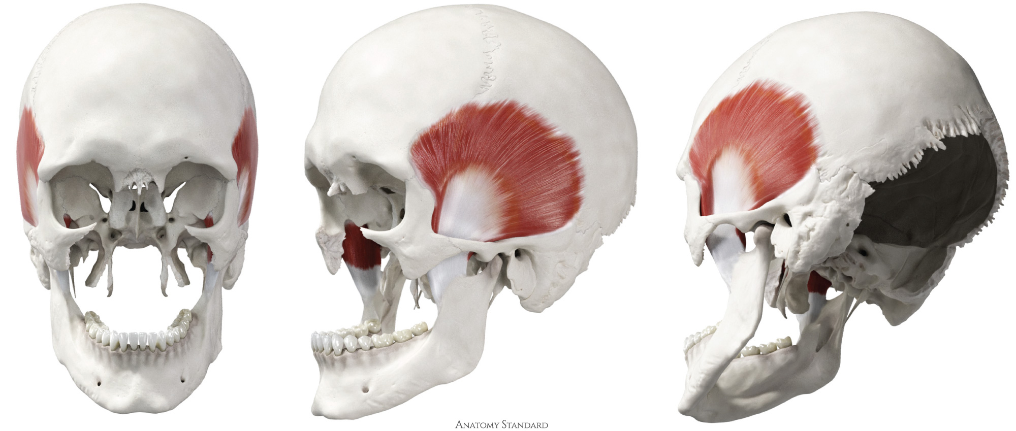
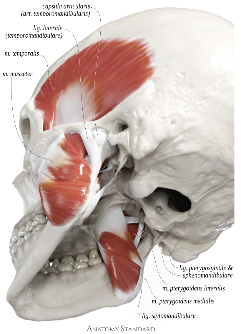
    Note the broad, circular, and complex-shaped connection of a muscle to the coronoid process of the mandible.*
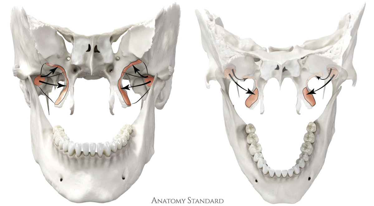
  • * Sun K, et al. Morphology of the temporalis muscle focusing on the tendinous attachment onto the coronoid process. Anat Cell Biol, 2021, 54(3):308–314.
  • Pterygoid Muscles


    Both pterygoid muscles, the medial and lateral, are indeed attached to the pterygoid process of the sphenoid bone. Due to their deep location and the less obvious spatial relationship between their heads, it can be challenging to correctly identify them.

    Icon of crossfade image The origins and insertions of the pterygoid muscles. Only skull bones that serve as the origin and insertion points of the pterygoid muscles are represented. Anterior oblique and posterior aspect.
    Medial and lateral pterygoid muscles with the related skull bones. Anterior oblique and posterior view Origins and insertions of the pterygoid muscles with the related skull bones. Anterior oblique and posterior view
    These images illustrates only the skull bones that serve as the origin or insertion points of the pterygoid muscles. The capusle of the TMJ is preserved. Anterior oblique & posterior aspect.
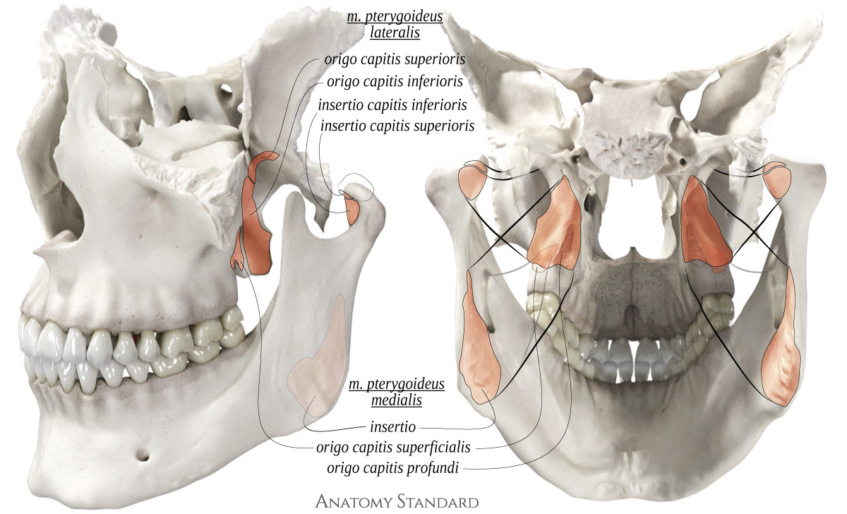
    Note the lower head of the lateral pterygoid passing in between the split heads of the medial pterygoid.

    Medial Pterygoid Muscle

    The fiber direction of the medial pterygoid muscle is similar to that of the masseter when observed from a lateral perspective. I use the 'M,' which is common for 'masseter' and 'medial,' as a mnemonic to differentiate the medial pterygoid from the lateral one.


    Icon of crossfade image Superficial and deep heads of the medial pterygoid muscle with the skull. Posterior ant lateral view
    Superficial and deep heads of the medial pterygoid muscle with the skull. Posterior ant lateral view Origins and insertion of the superficial and deep head of the medial pterygoid muscle with the skull. Posterior ant lateral view
    Overview of the medial pterygoid muscle. Posterior and lateral views. The posterior aspect shows only the skull bones that serve as origins or insertions for this muscle.
  • Origin: fossa pterygoidea (medial head) + tuber maxillae (lateral head)
  • Insertion: tuberositas pterygoidea maxillae
  • Function: elevatation of the mandible. It also contributes to limited protrusion. When acting unilaterally, it assists in moving the mandible to the contralateral side.

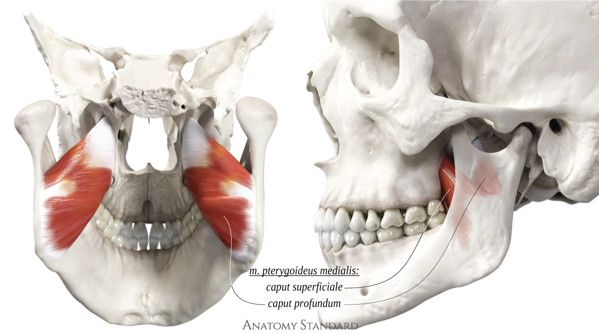
  • The medial pterygoid muscle has two heads: the superficial head is smaller and arises from the maxilla, while the deeper head is larger and originates from the pterygoid fossa.

    Lateral Pterygoid Muscle


    The lateral pterygoid is the smallest among all the masticatory muscles, yet it is the most complex. Notably, it is the only masticatory muscle with nearly horizontally aligned fibers. This muscle not only attaches to the bones but also to the capsule and articular disc of the TMJ. This is the only masticatory muscle with the almost horizontally aligned fibers. Due to its deep location in the infratemporal fossa and its surrounding tissue, anatomical dissection is very difficult.1, 2.

    Functionally and anatomically, the muscle is divided into two heads: upper and lower.
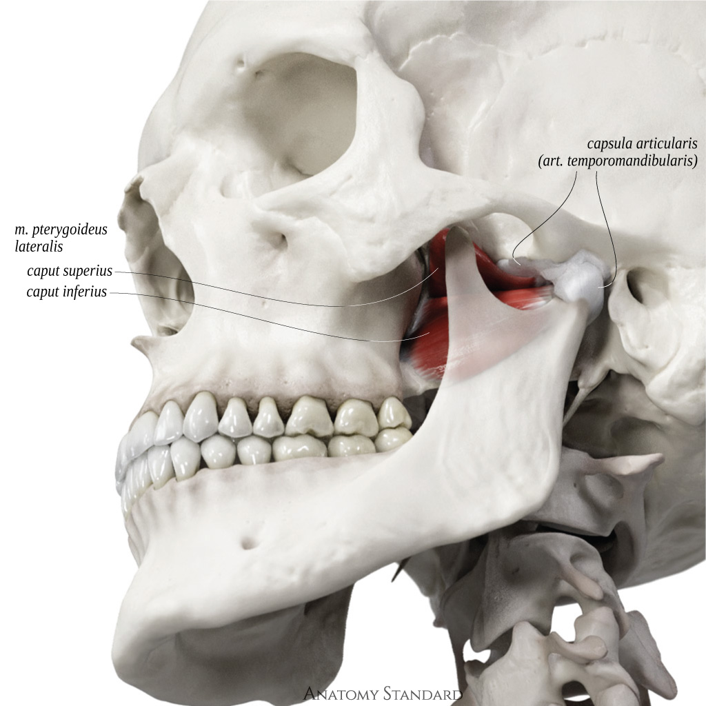

  • 1 Stöckle M, Fanghänel J et al. The morphological variations of the lateral pterygoid muscle: A systematic review. Ann Anat, 2018, 222:79–87.
  • 2 Haddioui A, Laison F, et al. Functional anatomy of the human lateral pterygoid muscle. Surg Radiol Anat, 2005, 27(4):271–286.
  • Icon of crossfade image Upper and lower heads of the lateral pterygoid muscle. Oblique  lateral aspect
    Upper and lower heads of the lateral pterygoid muscle. Oblique  lateral aspect Origin and insertion of the upper and lower head of the lateral pterygoid muscle. Oblique  lateral aspect
    Overview of the lateral pterygoid muscle
    Upper head:
  • Origin: infratemporal surface of the great wing of the sphenoid bone
  • Insertion: articular capsule of the TMJ and the TMJ articular disc
  • Function: pull the TMJ articular disc forward and medially
  • Lower head:
  • Origin: lateral surface of the lateral plate of the pterygoid process
  • Insertion: fovea mandibulae
  • Function: pull the condyle of mandible forward and medially
  • Electromyographic studies show that the heads of the muscle are active in different phases of opening and closing the lower jaw1. This data suggests the primary function of the upper head is to control the velocity at which the discocapsular system is pulled back into place during jaw closing movements, while the lower head pulls the mandible and discocapsular system forward during jaw opening movements2. This situation is quite unique, leading some scientists to consider the two heads of the lateral pterygoid as two functionally distinct muscles3.


  • 1 Hiraba K, Hibino K, et al. EMG activities of two heads of the human lateral pterygoid muscle in relation to mandibular condyle movement and biting force. J Neurophysiol, 2000, 83(4):2120–2137.
  • 2 Feneis H, Dauber W. Pocket atlas of human anatomy. 4th ed. Thieme, 2000.
  • 3 McNamara J. The independent functions of the two heads of the lateral pterygoid muscle. Am J Anat, 1973, 138(2):197–205.
  • Icon of crossfade image Origin and insertion of the upper and lower head of the lateral pterygoid muscle with the related skull bones. Anterior and oblique superior view
    Upper and lower head of the lateral pterygoid muscle with the related skull bones. Anterior and oblique superior view Origin and insertion of the upper and lower head of the lateral pterygoid muscle with the related skull bones. Anterior and oblique superior view
    The lateral pterygoid muscle and the related bones. Anterior and oblique superior aspect.
    The fibers of the lower head of the lateral pterygoid run obliquely. This means that when the muscles work bilaterally, they pull the condyles of the mandible forward, which is necessary for protrusion and depression movements of the mandible. However, in the case of unilateral action, the muscle significantly contributes to the lateral movement of the mandible in the contralateral direction. Simultaneously, the upper head adjusts the position of the articular disc in the temporomandibular joint. Any coordination issues between the heads may result in TMJ disorders, causing pain and limitation of the jaw movement, including partial or complete locking of the lower jaw1, 2, 3.
  • 1 Desmons S, Graux F, et al. The lateral pterygoid muscle, a heterogeneous unit implicated in temporomandibular disorder: a literature review. Cranio, 2007, 25(4):283–291.
  • 2 Писаревский ЮЛ, Найданова ИС, и др. Характеристика потенциала действия двигательных единиц латеральной крыловидной мышцы и биопотенциалов жевательных мышц при проведении сплинт-терапии по поводу болевой дисфункции височно-нижечелюстного сустава. Стоматология, 2019, 98(6):72–78.
  • 3 Murphy M, MacBarb R, et al. Temporomandibular disorders: a review of etiology, clinical management, and tissue engineering strategies. Int J Oral Maxillofac Implants, 2013, 28(6):e393–414.