Tarsus
The tarsal bones are quite different from the carpal bones of the hand. They are designed to physiologically distribute the body's weight to the plantar surface of the foot, absorb the shockwaves, and, finally, precisely measure the projection of the body mass center.


The list of terms:
TarsusMetatarsus
Ossa digitorum pedis – Phalanges of toes
Talus – Ankle bone
Calcaneus – Heel bone
Os naviculare – Navicular bone
Os cuboideum – Cuboid bone
Os cuneiforme laterale – Lateral cuneiform
Os cuneiforme intermedium – Intermediate cuneiform
Os cuneiforme mediale – Medial cuneiform
Talus
In contrast with the carpal bones, in the foot, there is only one bone directly articulating with the proximal extremity skeleton – only the talus articulates with the tibia & fibulatibia & fibula. The body weight pressure is distributed between the dorsally displaced tuber calcanei and – through the chain of other tarsal bones – to the metatarsal bones.
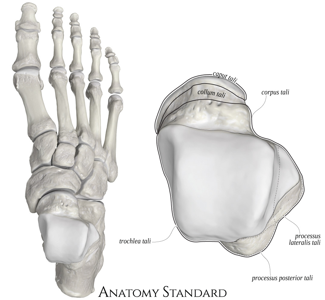


The main parts of the talus are the head (caput), neck (collum), and the body (corpus) that includes the large trochlea and two processes (lateral & posterior). The lateral process of the talus is the main gross asymmetrical structure that helps to distinguish the right talus from the left one.
The list of terms:
Caput tali – Head of talusCollum tali – Neck of talus
Corpus tali – Body of talus
Trochlea tali
Processus lateralis tali – Lateral process of talus
Processus posterior tali – Posterior process of talus
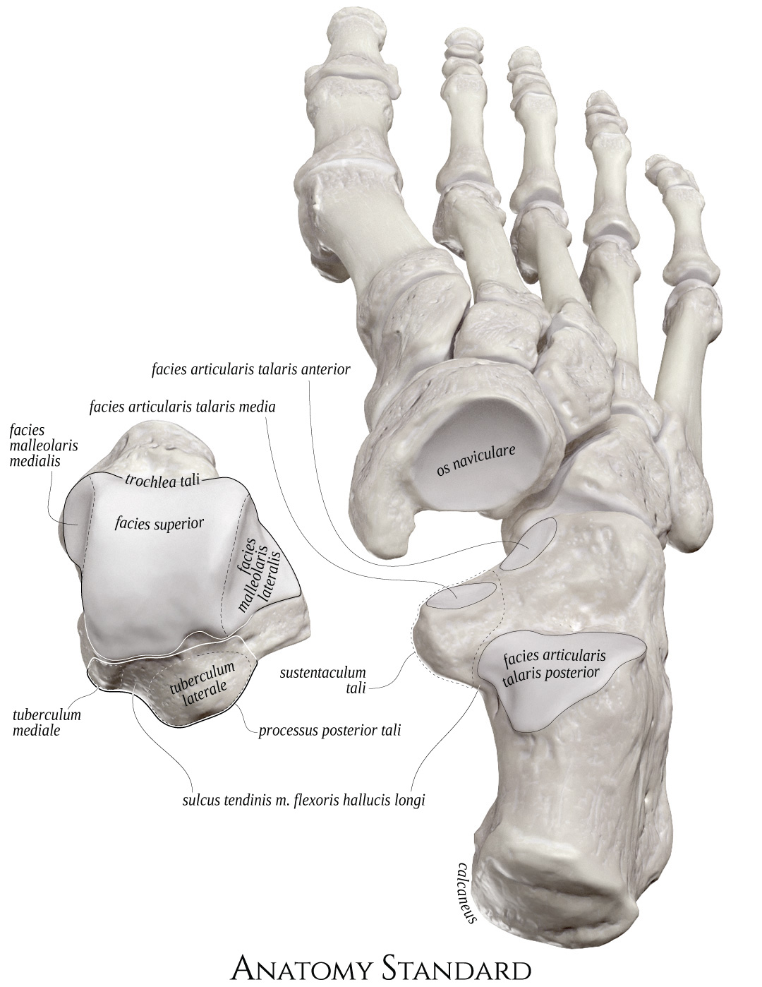


Click the image to see the bones directly contacting the talus – the concave surface of the navicular bone and three separate articulatory facets on the calcaneus's top surface.
The list of terms:
Trochlea taliFacies malleolaris medialis – Medial malleolar surface
Facies superior – Superior surface
Facies malleolaris lateralis – Lateral malleolar surface
Processus posterior tali – Posterior process of talus
Tuberculum mediale – Medial tubercle
Tuberculum laterale – Lateral tubercle
Sulcus tendinis m. flexoris hallucis longi – Groove for the tendon of flexor hallucis longus muscle
Os naviculare – Navicular bone
Calcaneus – Heel bone
Facies articularis talaris anterior – Anterior facet for the talus
Facies articularis talaris media – Middle facet for the talus
Facies articularis talaris posterior – Posterior facet for the talus
Sustentaculum tali


Please note that in contrast with the three separate articular facets on the calcaneus's top surface, the talus's congruent anterior and middle articular facets are usually fused with the head of the talus.
The list of terms:
Caput tali – Head of talusFacies articular navicularis – Navicular articular surface
Facies articulars calcanea anterior – Anterior calcanean facet
Facies articularis calcanea media – Middle calcanean facet
Facies articularis calcanea posterior – Posterior calcanean facet
Sulcus tali – Sulcus of talus
Sulcus tendinis musculi flexoris hallucis longi – Groove for the tendon of m. flexor hallucis longus
Calcaneus


The most prominent feature seen from the medial aspect of the heel bone is the sustentaculum tali. The word sustentaculum derives from the Latin and means support or sustenance. That term seems to be appropriate: the pressure transmitted through the talus (i.e., the weight of the whole body) is, to a great extent, directed toward that medial prolongation of the heel bone.
The list of terms:
Tuber calcaneiProcessus lateralis tuberis calcanei – Lateral process of calcaneus
Processus medialis tuberis calcanei – Medial process of calcaneus
Tuberculum calcanei – Calcaneal tubercle
Sustentaculum tali
Sulcus tendinis m. flexoris hallucis longi – Groove for the tendon of m. flexor hallucis longus
Facies articularis cuboidea – Cuboid articular surface
Facies articularis talaris anterior – Anterior facet for the talus
Facies articularis talaris media – Middle facet for the talus
Facies articularis talaris posterior – Posterior facet for the talus


You will find the three distinct articulatory facets for talus bone on the superior surface of the calcaneus. The lateral facet localizes on top of the sustentaculum.
The list of terms:
Sustentaculum taliSulcus tendinis m. flexoris hallucis longi – Groove for the tendon of m. flexor hallucis longus
Facies articularis cuboidea – Cuboid articular surface
Facies articularis talaris anterior – Anterior facet for the talus
Facies articularis talaris media – Middle facet for the talus
Facies articularis talaris posterior – Posterior facet for the talus
Sulcus calcanei
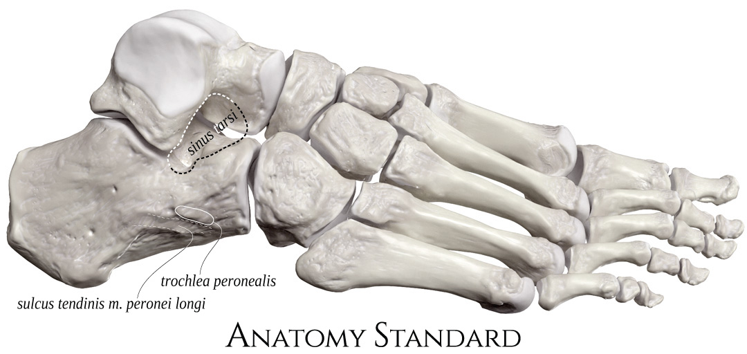


The list of terms:
Sinus tarsiTrochlea peronealis – Peroneal trochlea
Sulcus tendinis m. peronei longi – Groove for the tendon of the peroneus longus muscle
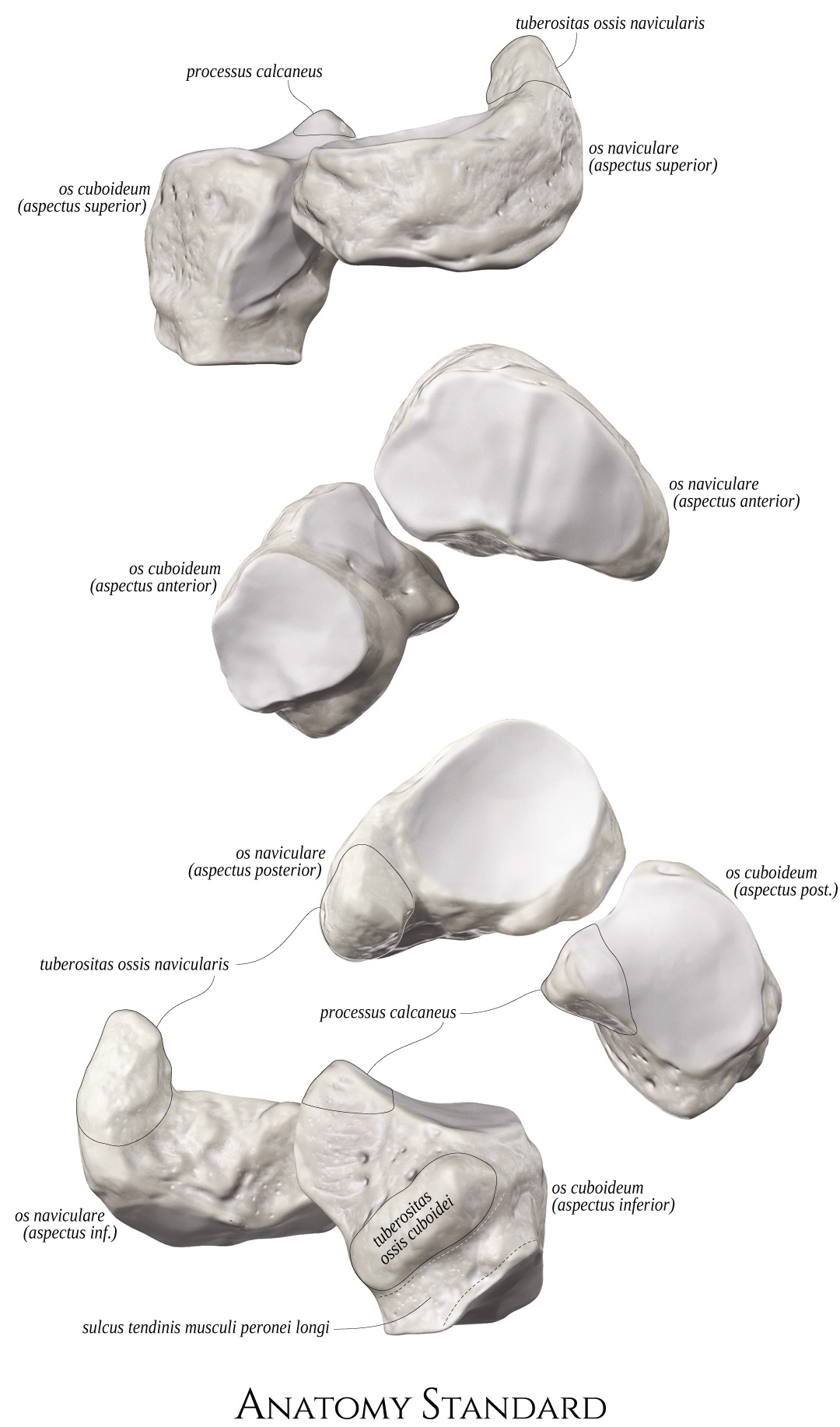
Os Naviculare & Os Cuboideum


Click an image to see the foot's functional split: the navicular bone transmit the pressure force from the talus to the cuneiform bones and further – to the 1st, 2nd, and 3rd metatarsal bone. In contrast, the cuboid bone transmits the calcaneus's pressure force directly to the 4th and 5th metatarsal bones.


The list of terms:
Tuberositas ossis navicularis – Tuberositty of navicular boneProcessus calcaneus – Calcanean process
Tuberositas ossis cuboidei – Tuberosity of cuboid
Sulcus tendinis m. peronei longi – Groove for tendon of peroneus longus muscle
Os Cuneiforme Mediale, Intermedium & Laterale
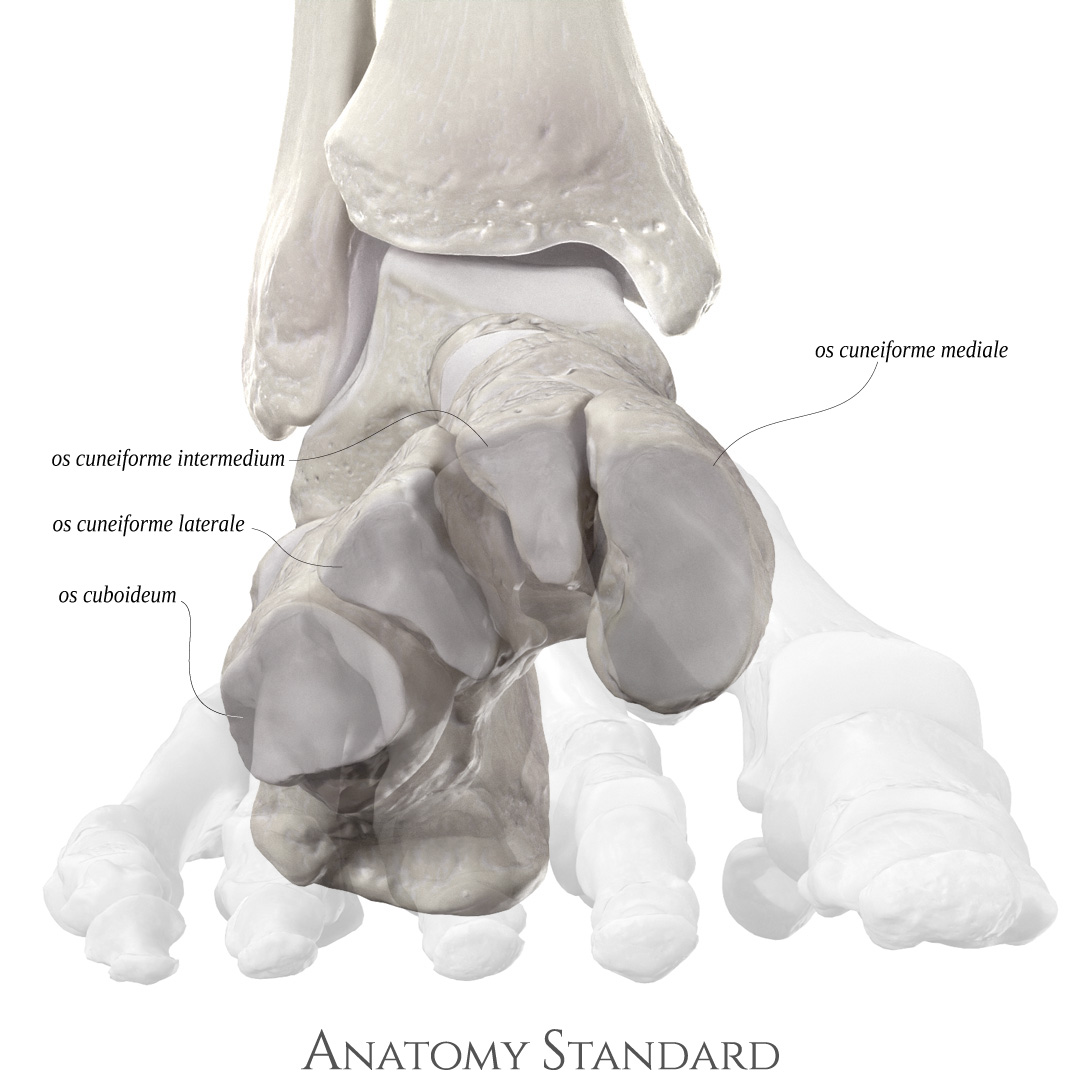


Note the wedge-like shape of these bones (from Latin cuneus – wedge).
The list of terms:
Os cuboideum – Cuboid boneOs cuneiforme mediale – Medial cuneiform
Os cuneiforme intermedium – Intermeiate cuneiform
Os cuneiforme laterale – Lateral cuneiform


The list of terms:
Os cuneiforme mediale – Medial cuneiformOs cuneiforme intermedium – Intermeiate cuneiform
Os cuneiforme laterale – Lateral cuneiform
Last update: 20/Dec/2020